


They’re tiny, transparent, and often the last surviving fish left swimming in your aquarium. And they’re quickly taking over laboratories around the world as the most popular test animal. Goodbye lab rat, hello zebrafish.
It wasn’t until the early ’70s that scientists placed the common zebrafish under the microscope and discovered their transparent skin allowed easy access to observing organ growth. As vertebrates, zebrafish embryos develop through a similar process as human embryos. They have many of the same organ systems, including a spinal cord, muscles, a heart, and blood. Now a team of Berkeley scientists studying hearing loss is looking deeper into zebrafish anatomy for answers—into the inner ear.
Humans are born with 16,000 hair cells—called stereocilia—in the inner ear, which respond to vibrations to help us keep our balance and determine where a sound is coming from. Soft noises cause these hair cells to shift at their bases; loud noises can cause them to break. And once they’re gone, they never grow back in humans. Zebrafish, however, regenerate stereocilia all the time.
Dr. Manfred Auer, a staff scientist with the Lawrence Berkeley National Laboratory’s Life Sciences Division, wants to know why, and how. He’s leading a study to find out which proteins exist in the inner ears of zebrafish that allow them to regain lost hearing. It is a complex and delicate search, sifting through about 30,000 different proteins, but one that has incredible potential.
“More than the loss of sight, the loss of hearing is often described as the most debilitating,” Auer says. One out of a thousand children in the United States is born deaf, and 30 million Americans develop hearing loss during their lifetime. Many have learned to adapt to hearing loss with hearing aids and sign language, but the possibility of actually restoring lost hearing is what gets Auer really excited. He describes his quest as nothing less than the “Holy Grail of research.”
But it wasn’t until a new technique in electron microscope imaging was developed that scientists could actually get a good look at the hair cells in the inner ears of zebrafish embryos, which are themselves the size of a pinhead.
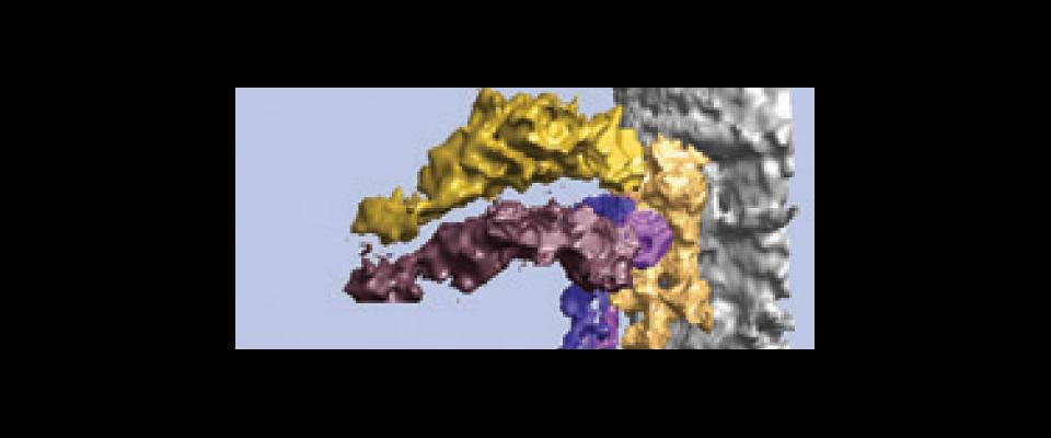
With electron microscope tomography, Auer and his team created three-dimensional images of zebrafish hair cells by tilting the slide under the electron beam at 140 different angles. Auer says studying transparent zebrafish with this “very fancy tool” can advance research on many human health problems. “There are no limitations—from breast cancer to kidneys to bacterial colonies,” Auer says. “We’re going beyond what’s been possible before because we’ve added a third dimension.”
From the May June 2006 What’s Happened to the Animals of Yosemite issue of California.